Free science diagrams to download
Labelled and unlabelled biology diagrams with printable worksheets and answer keys: plant cell, animal cell, cell membrane, mitochondria, mitosis, meiosis, cell cycle, ear, digestive system, heart, human eye, neuron, microscope, water cycle, photosynthesis, dna structure, bacterial cell, virus. Download SVG, PNG or PDF free under CC BY 4.0, or open any diagram in LabCanvas to edit the colours and labels.
Last updated
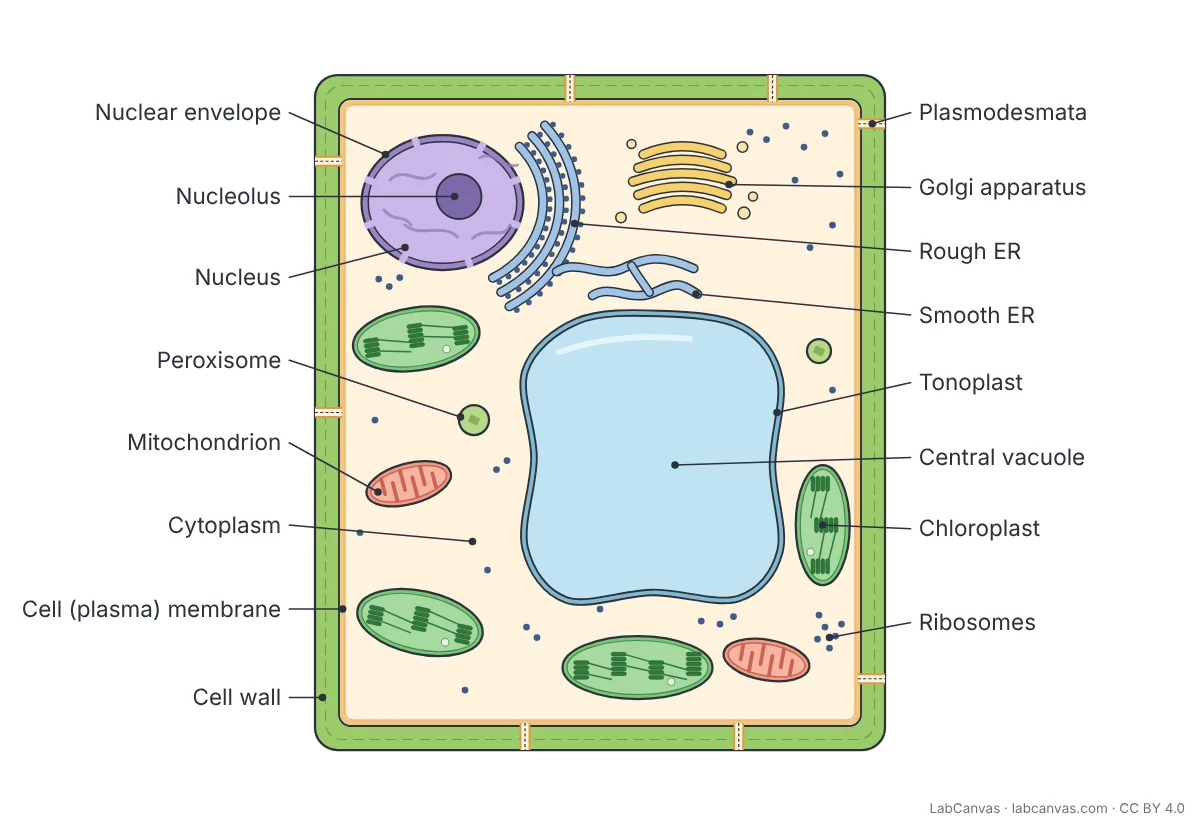 Plant cell diagram
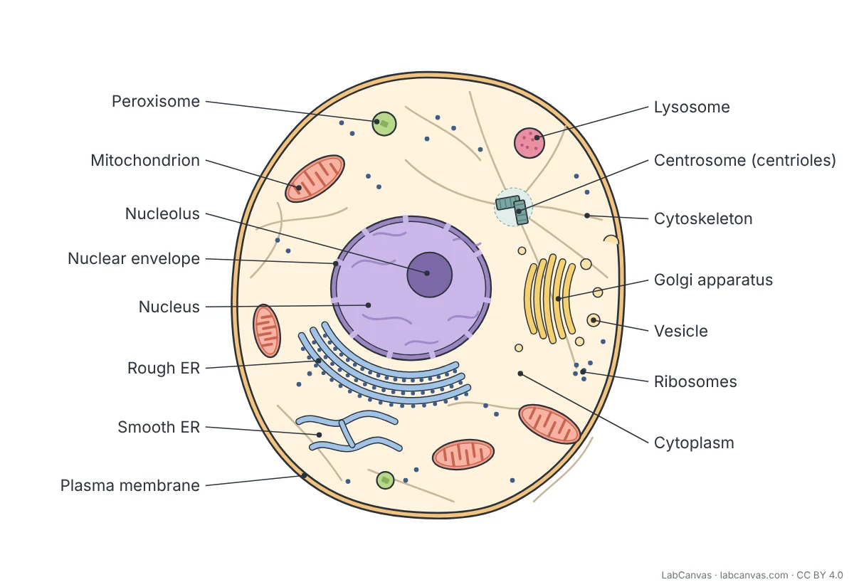 Animal cell diagram
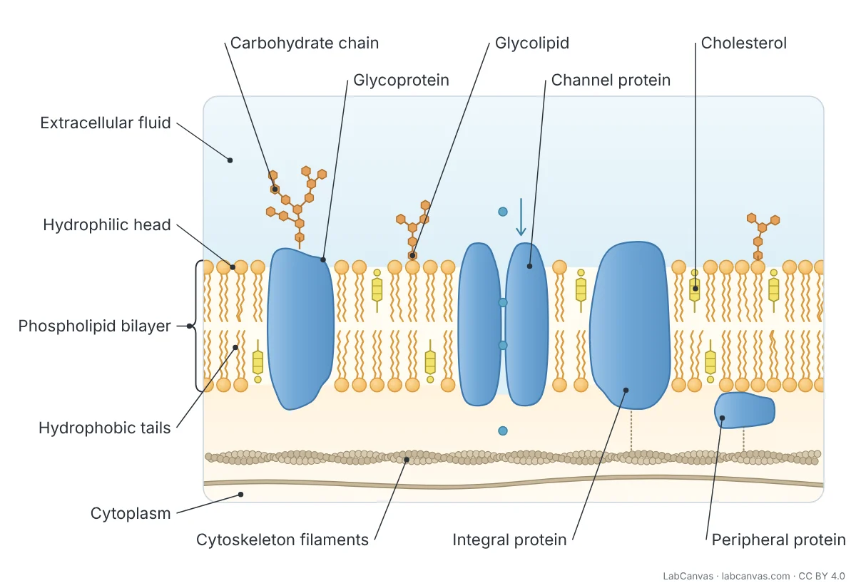 Cell membrane diagram
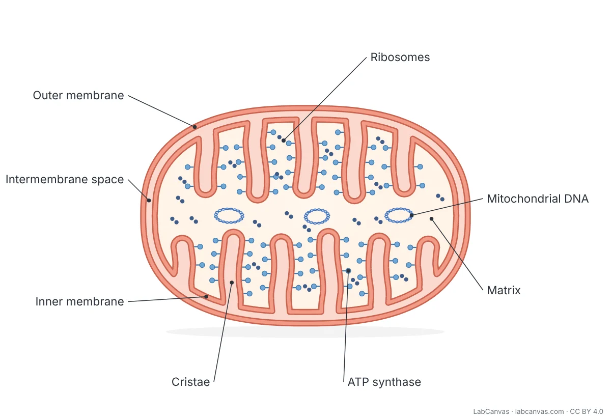 Mitochondria diagram
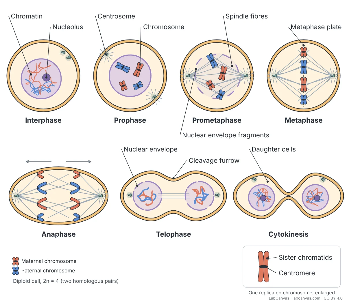 Mitosis diagram
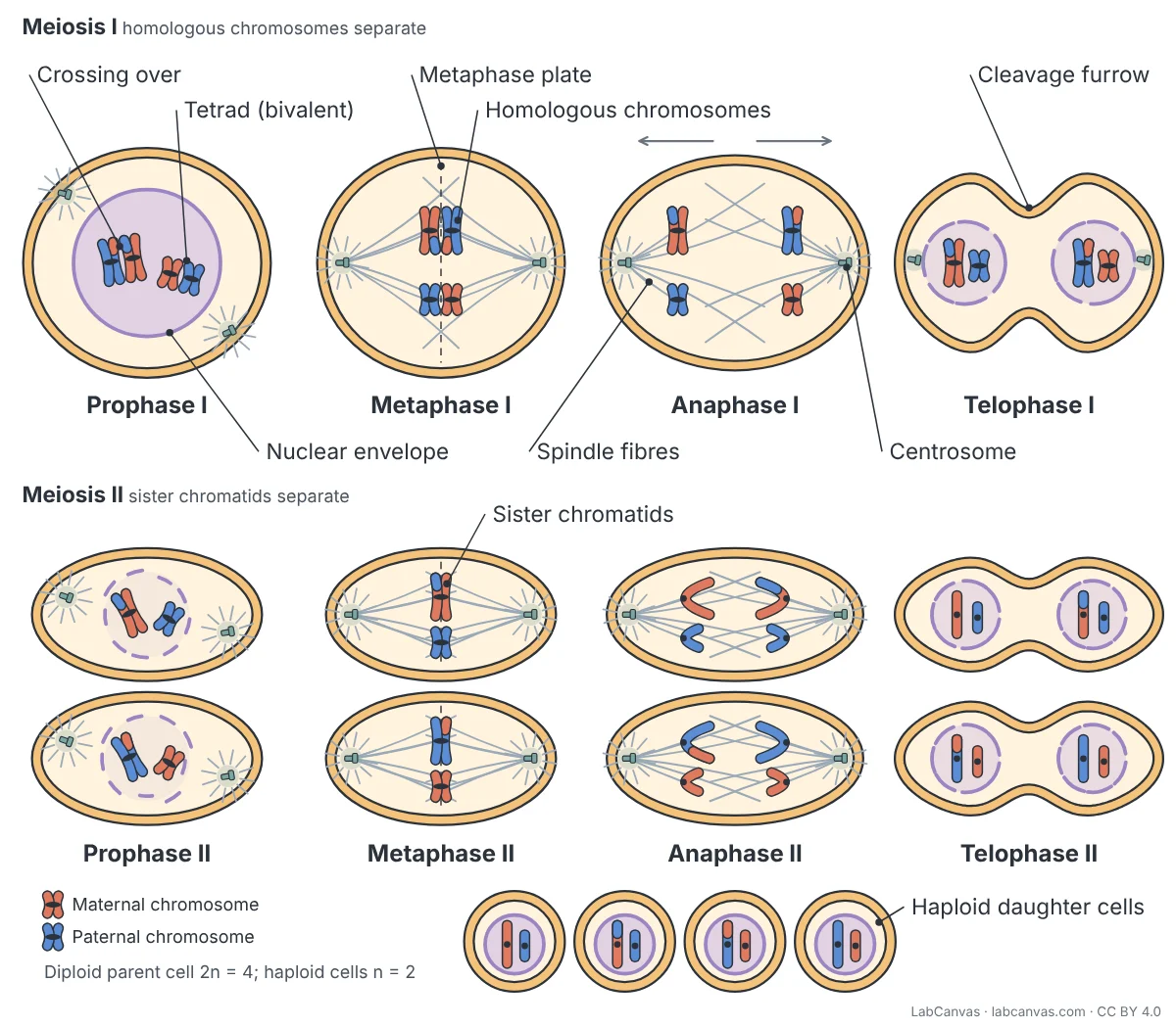 Meiosis diagram
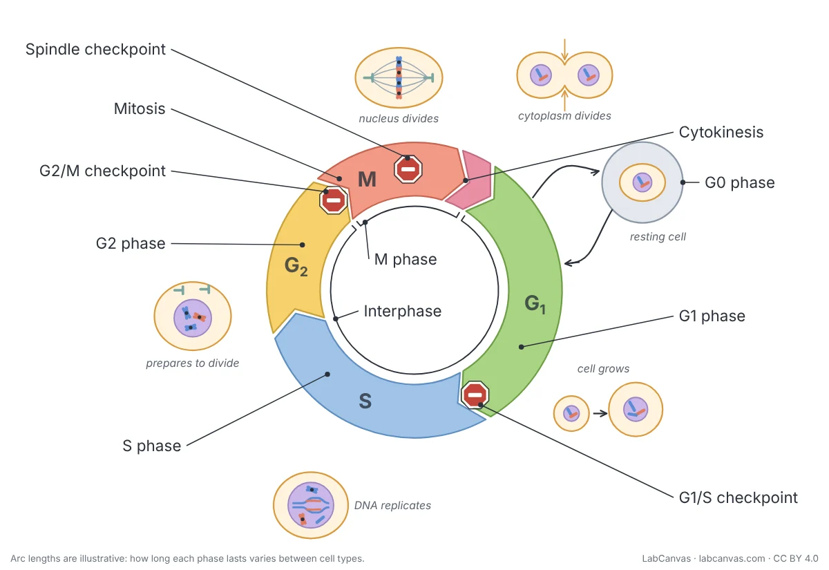 Cell cycle diagram
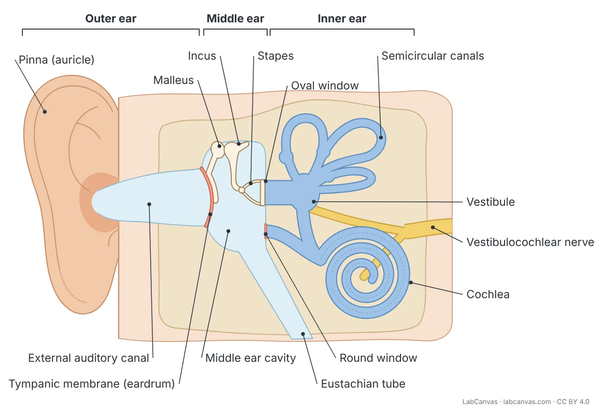 Ear diagram
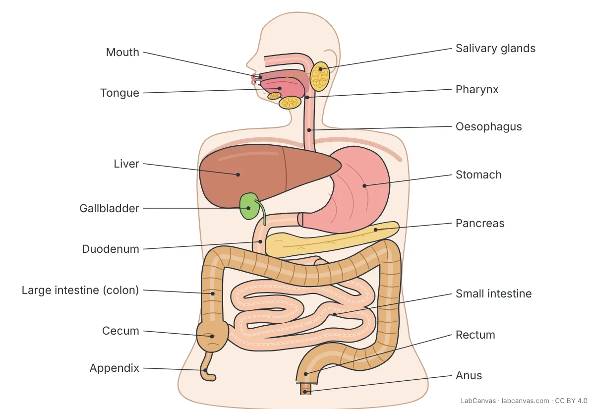 Digestive system diagram
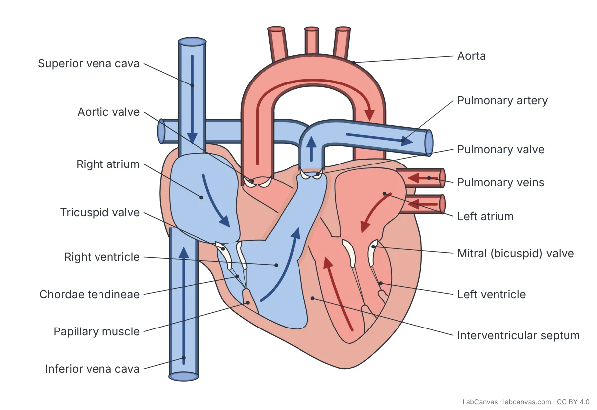 Heart diagram
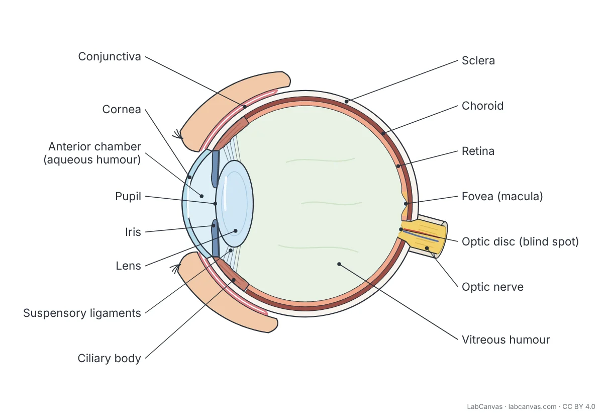 Human eye diagram
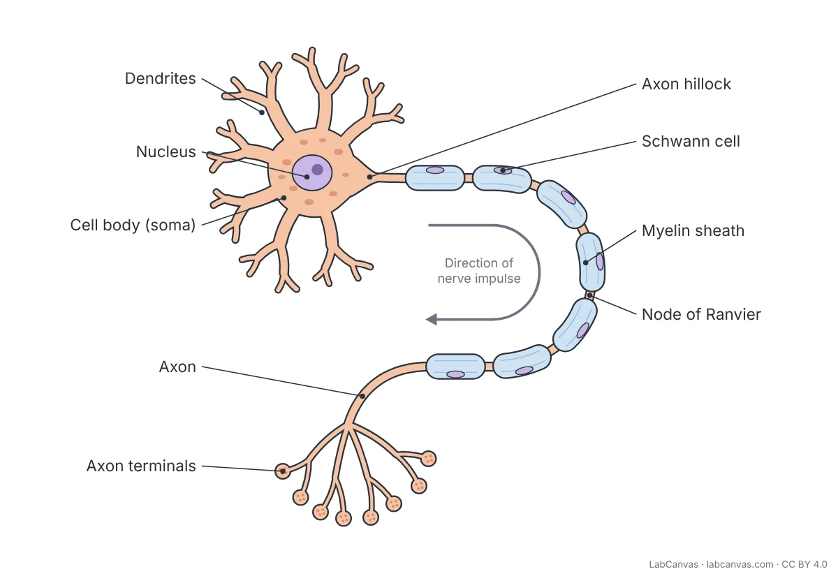 Neuron diagram
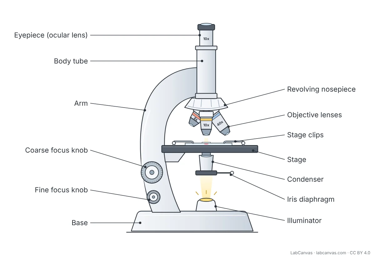 Microscope diagram
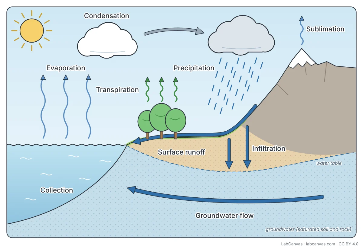 Water cycle diagram
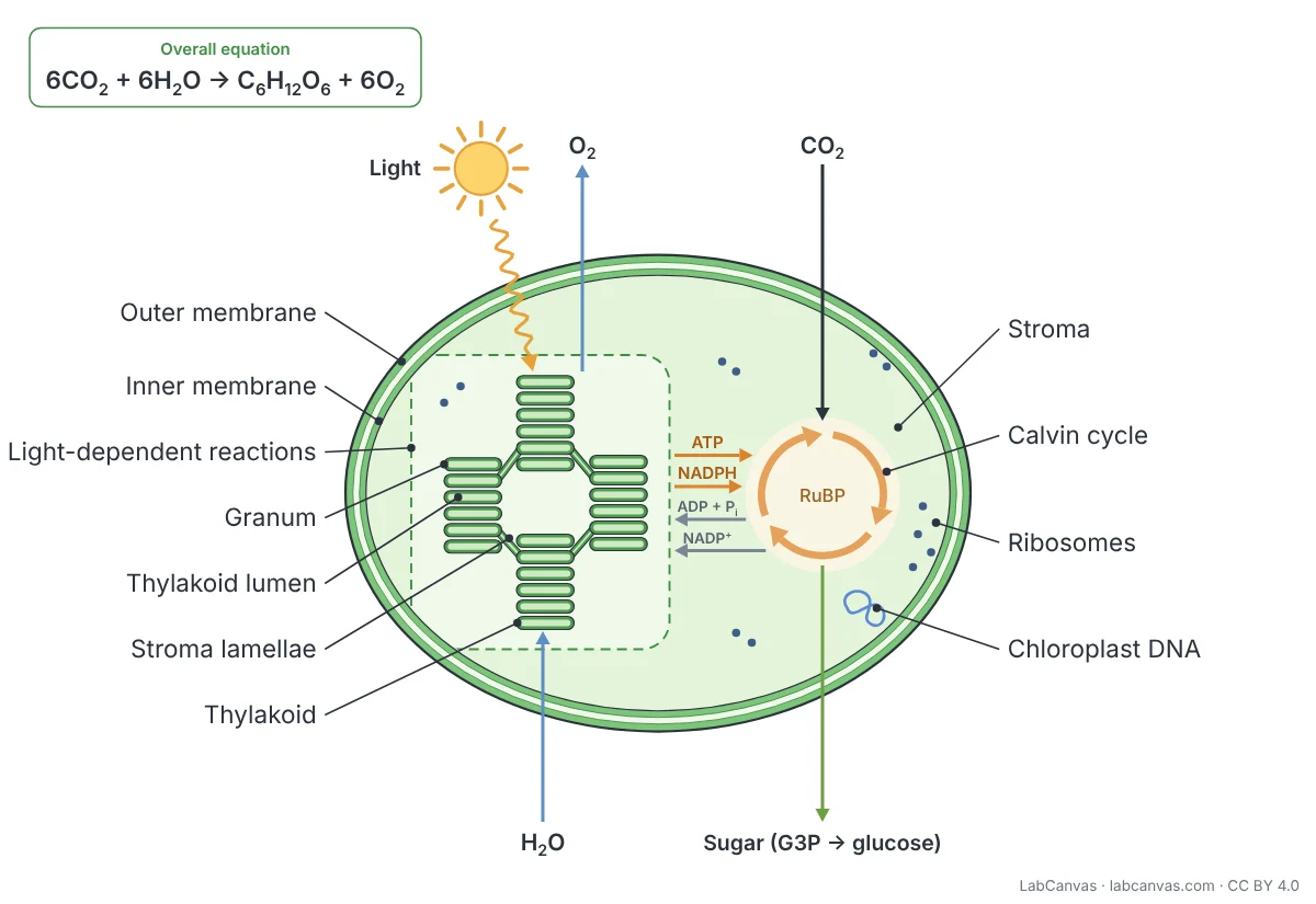 Photosynthesis diagram
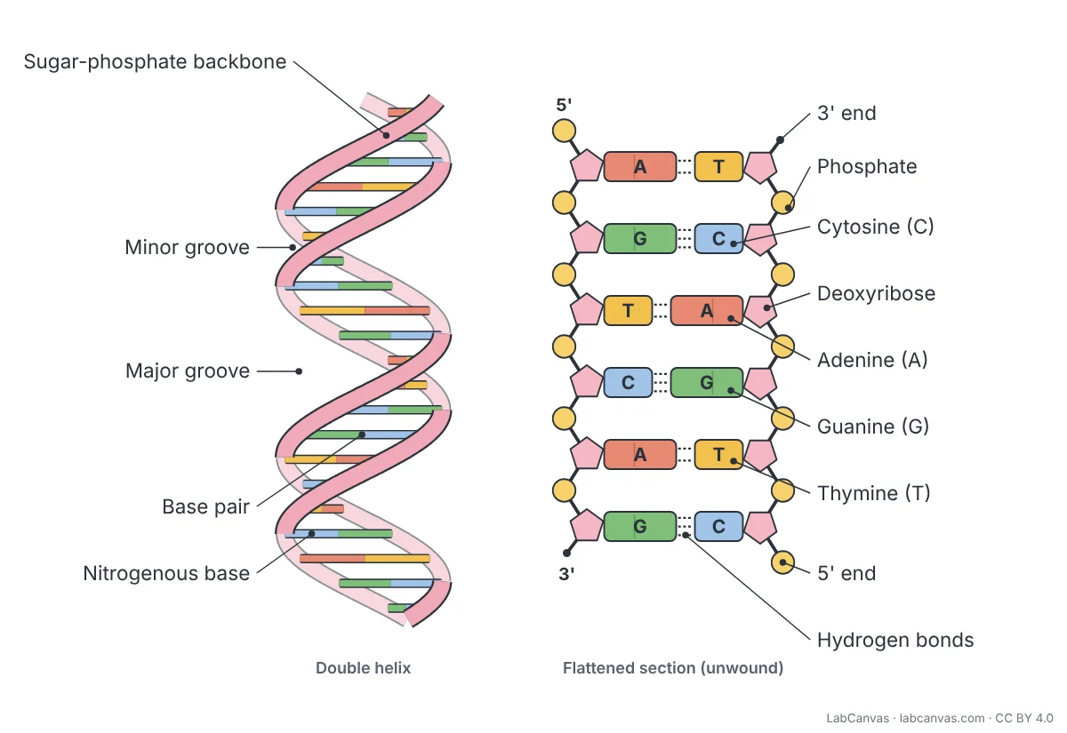 DNA structure diagram
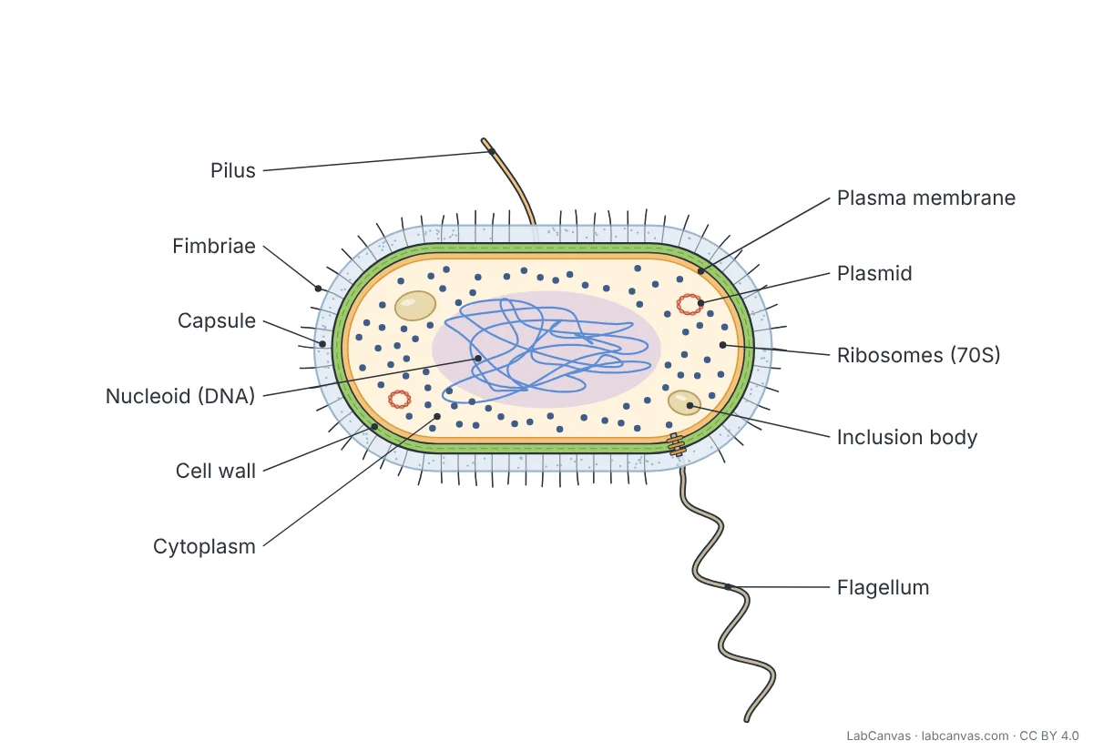 Bacterial cell diagram
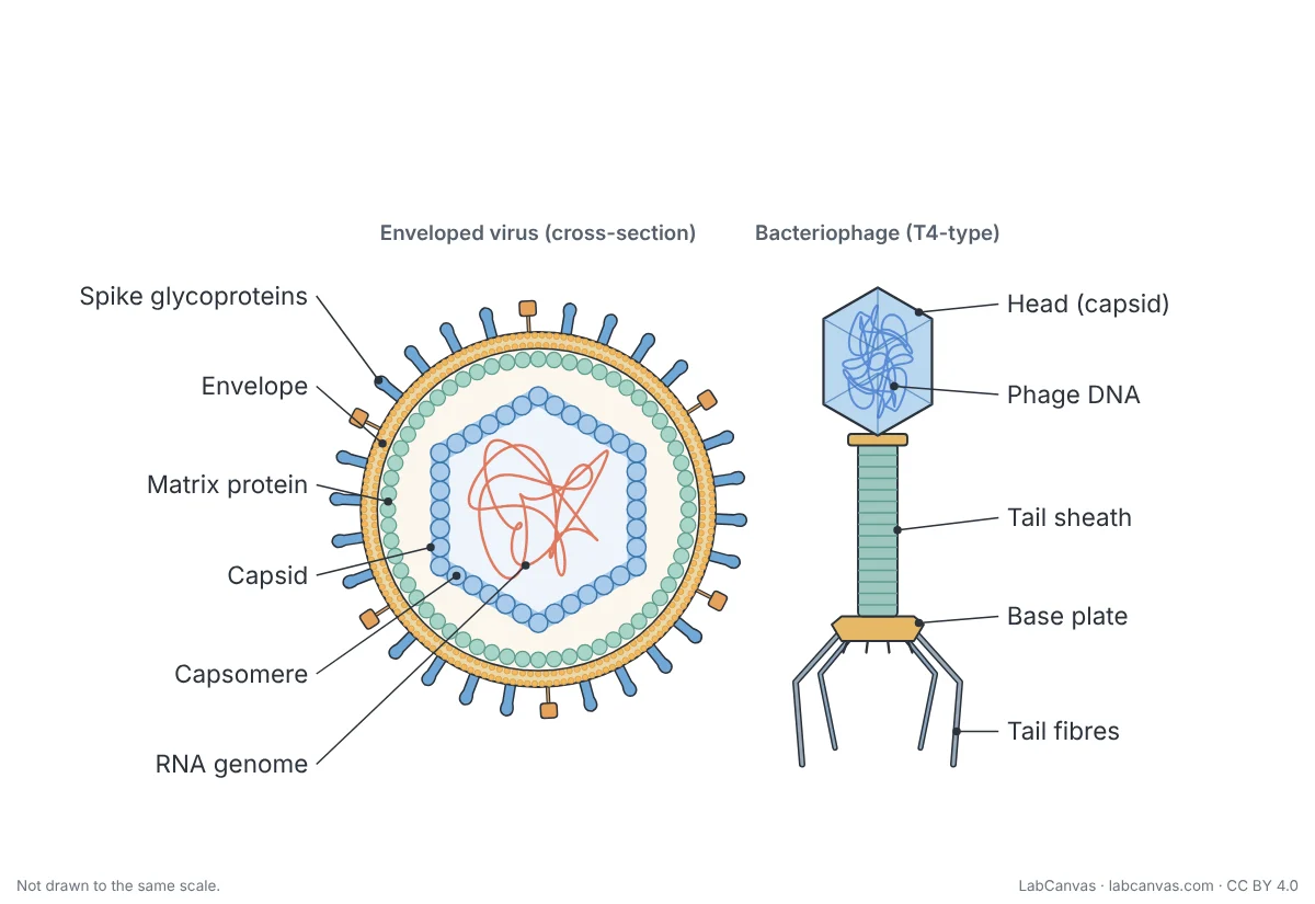 Virus diagram
What each diagram page includes
Every diagram has its own page, and every page is built the same way, so once you know one you know them all. At the top is the diagram in three versions you can switch between: labelled, with every part named; unlabelled, the same drawing with the labels removed; and a worksheet, with numbered blanks in place of the labels. Below it are the downloads: the labelled and unlabelled diagrams as SVG (vector, sharp at any size) and as 3200-pixel PNG with a transparent background, the worksheet as PNG, and a printable A4 PDF with the worksheet and its answer key.
Under the downloads, a table lists every labelled part with a one-line description of what it is or does, numbered in the same order as the blanks on the worksheet, so the table doubles as the answer key on screen. Each page then explains how to label the diagram step by step, compares it with a related structure or process in a table (plant against animal cells, mitosis against meiosis, and so on), adds extra explanation where it helps, and ends with a five-question quiz with the answers explained, frequently asked questions and the textbook sources for every fact.
Diagrams by topic
- Cells and their parts: animal cell, plant cell, bacterial cell, cell membrane, mitochondria, virus.
- Cell division and DNA: cell cycle, mitosis, meiosis, dna structure.
- The human body: heart, human eye, ear, neuron, digestive system.
- Processes and the lab: photosynthesis, water cycle, microscope.
All of them are also listed with pictures in the Education section of the blog. For research figures rather than teaching diagrams, see the figure templates, which are drawn for papers and cite the studies they follow.
Using the diagrams in class and for revision
For teaching, project the labelled diagram while you explain each part, then show the unlabelled version and ask the class to name the parts. The A4 worksheet PDF has the numbered diagram with lines to write on, and its answer key on the second page, so it works as homework, a starter activity or a quick test. For revision, cover the parts table, label the worksheet from memory and then check it against the table, and use the quiz at the end of the page to test the ideas behind the labels, not only the names. The SVG and PNG files drop straight into slides, handouts, posters and learning platforms; the transparent PNG sits cleanly on coloured backgrounds.
Editing a diagram
Every diagram opens in the LabCanvas figure editor in your browser: click Edit it in LabCanvas on its page. It opens as separate shapes and text rather than a flat picture, so you can recolour parts to match your slides, translate or simplify the labels, remove parts your course does not cover, add arrows and notes, or combine two diagrams into one figure. You can then export the result as SVG, PNG or PDF at the size you need.
How to credit or cite a diagram
The diagrams are free under the Creative Commons Attribution 4.0 licence (CC BY 4.0): you may copy, adapt and share them, including commercially and in publications, as long as you give credit. Each page shows a ready-made credit line to copy, in the form “Heart diagram by LabCanvas (labcanvas.com), CC BY 4.0”. Put it in the figure caption, on the slide or in the image credits. If you changed the diagram, say so, for example “adapted from”. In a thesis or report, cite the diagram page’s web address and the date you downloaded it, as you would any other online figure.
How the diagrams are checked
The labels and descriptions follow open, peer-reviewed introductory textbooks, mostly OpenStax Biology 2e and Anatomy and Physiology 2e, and each page lists the chapters it used. The drawings are simplified teaching illustrations: they show the parts a school or first-year university course expects, in a clear arrangement, and they are not drawn to scale.
Questions about the diagrams
Are the diagrams free to use?
Yes. Every diagram is free to download and use, including in homework, teaching materials, slides, posters, websites and publications, under the Creative Commons Attribution 4.0 licence (CC BY 4.0). Credit “LabCanvas (labcanvas.com)”.
Which formats can I download?
Each diagram comes as SVG (vector), as a 3200-pixel PNG with a transparent background, labelled and unlabelled, and as a printable A4 worksheet with numbered blanks and an answer key (PDF).
Can I change the labels or colours?
Yes. Click “Edit it in LabCanvas” on a diagram page: it opens in the LabCanvas figure editor as separate shapes and text you can recolour, move and relabel.
Are the diagrams accurate?
They follow the OpenStax Biology 2e and Anatomy and Physiology 2e textbooks, which each page cites. They are simplified teaching illustrations, not drawn to scale.