The LabCanvas 3D Studio: adjustable 3D models of cells, nanomaterials and devices
How to make 3D scientific renders in the LabCanvas 3D Studio: adjustable models of cells, viruses, nanoparticles, 2D materials, MOFs and devices, molecules from PubChem, proteins from the PDB, crystals from CIF files, and renders placed in figures.
Contents
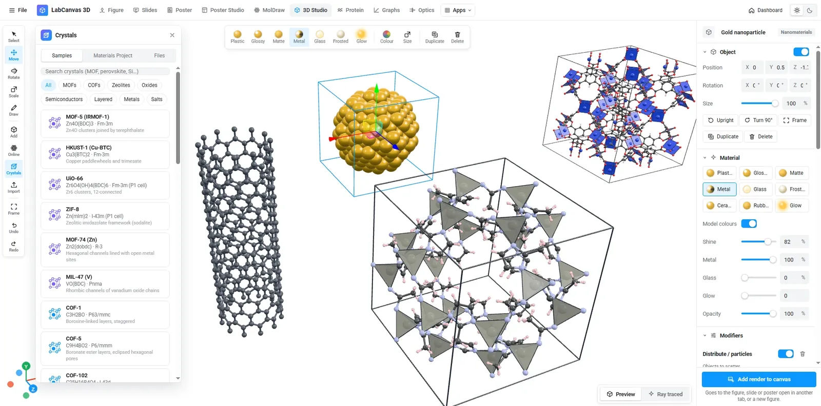
The LabCanvas 3D Studio makes 3D pictures for science without 3D modelling. Its models are generators: a gold nanoparticle, a liposome, a virus, a cell cut open, a MOF or a battery stack is built from settings you change with sliders. Molecules come from PubChem, proteins from the RCSB PDB and crystals from CIF and POSCAR files. Renders go straight into your figures.
Models by subject
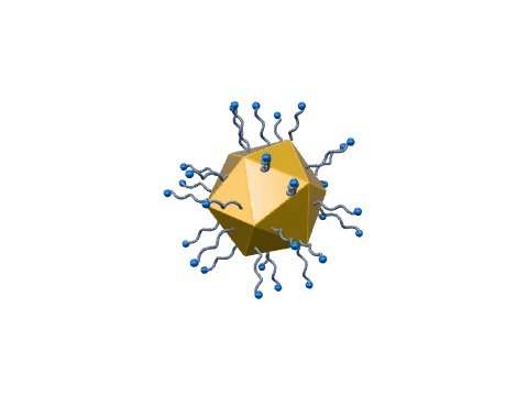
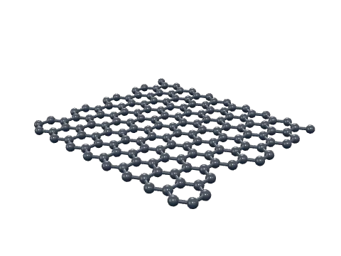
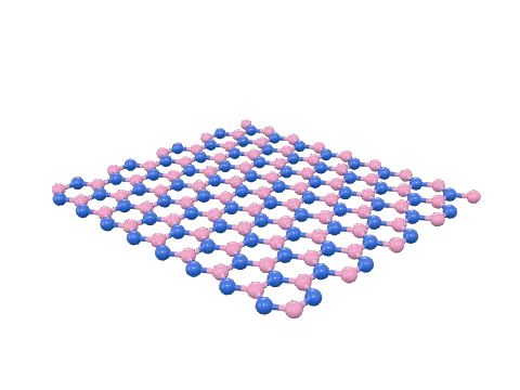
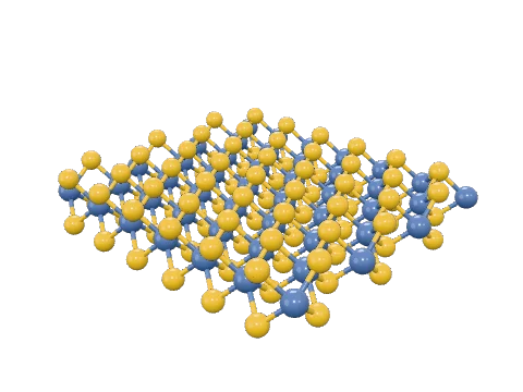
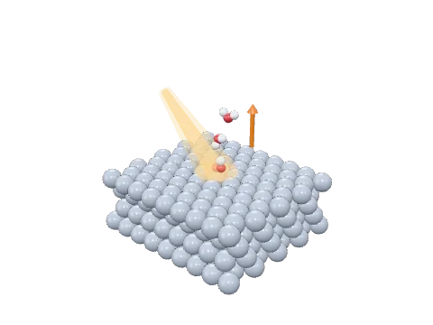
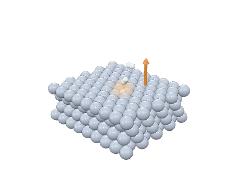
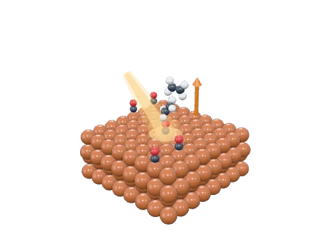
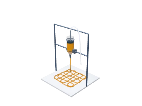
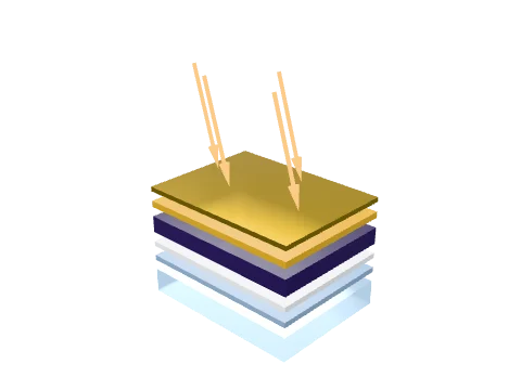
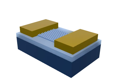
The Add menu groups the models by subject, each with a small picture so you can see what you add. Here are a few from each group.
Biology
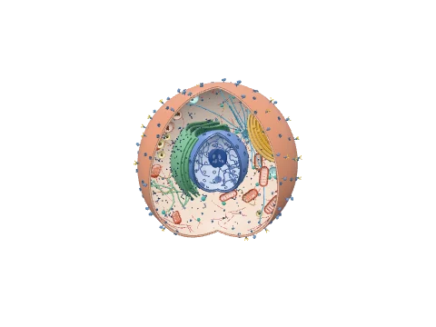
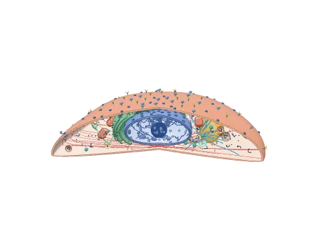
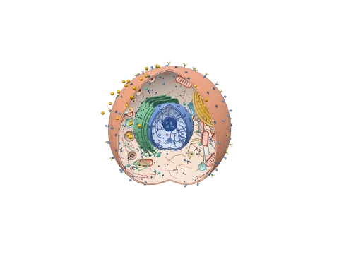
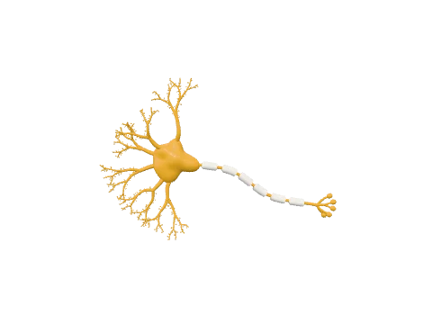
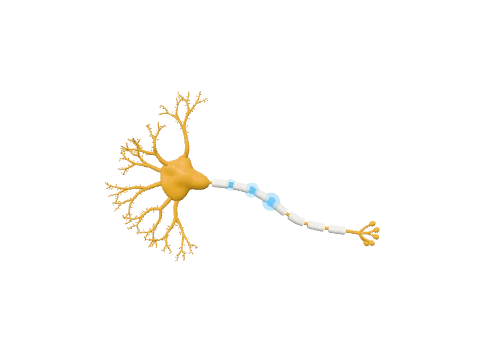
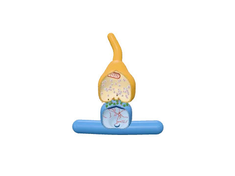
Nanomaterials
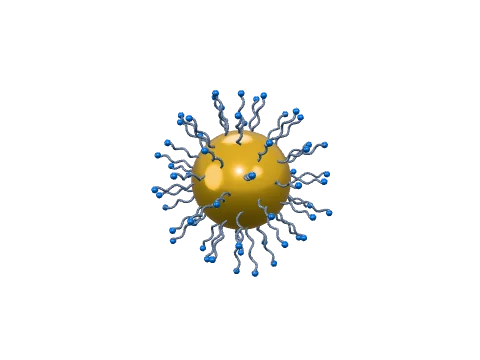
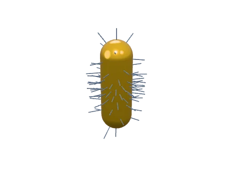
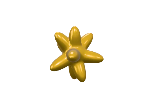
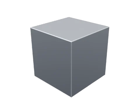
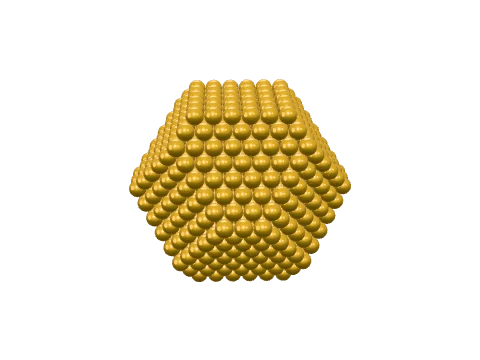
2D materials, catalysis and devices
What the 3D Studio can do
| Feature | Details |
|---|---|
| Adjustable models | Cells and organelles, viruses, antibodies, liposomes and LNPs, nanoparticles atom by atom, quantum dots, 2D materials, MOFs, zeolites, scaffolds, devices and shapes |
| Molecules and proteins | PubChem compounds and RCSB PDB structures, or your own PDB, mmCIF, SDF and XYZ files |
| Crystals | CIF and POSCAR files, Materials Project search, unit cells and supercells, ball-and-stick, space-filling or polyhedra |
| Molecule brushes | DNA, RNA, chromatin, helices, strands, collagen, actin, microtubules and polymer chains along a freehand stroke |
| Modifiers | Array, displace, distribute and gradient |
| Output | PNG at 1×, 2× or 4×, GLB, or a render placed straight into a figure |
How to make a 3D render for a figure
- Open the 3D Studio and click Add.
- Pick a model, for example Nanomaterials › Gold nanoparticle, and drag it into the view.
- Change its shape, size and surface in the panel on the right.
- Choose a lighting preset and a background (white, a colour or transparent).
- Click Add render to canvas to send it to your figure, or export a PNG.
Loading only what you use
The 3D Studio has hundreds of models, but it loads the code of a model only when you add it. The Add menu shows a small picture of every model, so you see the whole library without waiting for any of it to load.
Frequently asked questions
Do I need to know Blender to use the 3D Studio?
No. Models are made by generators with sliders: pick a nanoparticle, a cell or a crystal from the Add menu and change its settings in the panel. Move, rotate and scale with the toolbar or the W, E and R keys.
Can I open my own 3D files?
Yes: GLB, OBJ, STL, PLY, FBX, 3MF, PDB, mmCIF, SDF, MOL and XYZ, plus CIF and POSCAR crystal files.
How do I get a crystal structure?
Open a CIF or POSCAR file, search the Materials Project, or pick a ready-made MOF, COF, zeolite or mineral in the Crystals panel. Show it as ball-and-stick, space-filling or polyhedra, as a unit cell or a supercell.
How do I put a render in my figure?
Click Add render to canvas. The render goes into the figure, slide or poster open in another tab, or into a new figure. You can also export a PNG at 1×, 2× or 4×.
Does it run on a laptop?
Yes. The 3D Studio runs in the browser with WebGL; only the model you add is loaded, so the app starts quickly. Ray traced renders take longer on slower graphics hardware.
Try it in LabCanvas
Every app is free to start in your browser. Create an account and begin.
Start 3D Studio free in your browserKeep reading

ImageJ vs LabCanvas Image Analysis: an honest comparison
ImageJ/Fiji or LabCanvas Image Analysis? Six everyday jobs done in both, six newer ones (macros, vendor files, 3D, StarDist, big images) and which to use.
LabCanvas Image Analysis: measure microscopy images, western blots and gels in your browser
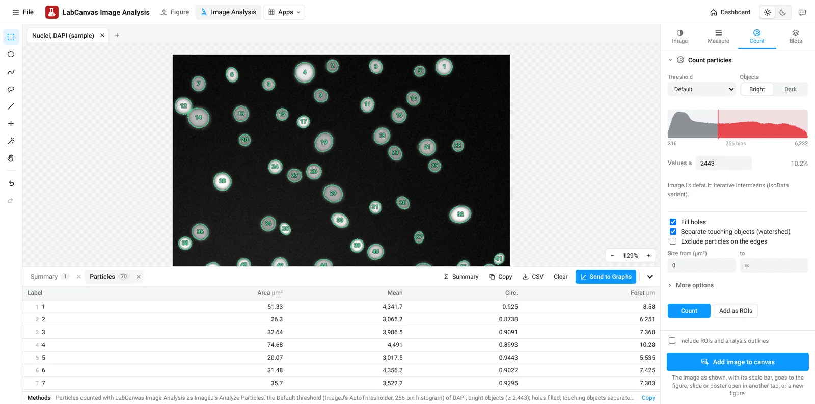
How to measure images in LabCanvas Image Analysis: guided assays (nuclear translocation, spots per cell, wound healing, FRAP, calcium, tracking, branching), western blots with a ladder and total-protein normalisation, cell counting with a trainable classifier, ROIs with ImageJ definitions, on the raw pixel values, with results sent to Graphs.
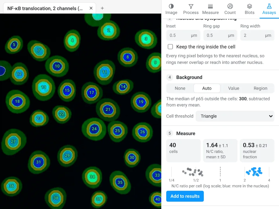
Image Analysis in LabCanvas: a complete guide to every tool and setting, with examples
Every tool in LabCanvas Image Analysis, tab by tab, with its settings and a real example: image check, display and scale, stacks and Z projection, background subtraction, filters, thresholds and binary operations, ROIs and Set measurements, Analyze Particles, a pixel classifier, spots, cell counter, western blot densitometry, colocalisation, colour deconvolution and eleven guided assays.