How to cite ImageJ and Fiji (with a methods sentence to copy)
Cite ImageJ (Schneider et al. 2012), Fiji (Schindelin et al. 2012) or ImageJ2 (Rueden et al. 2017): DOIs, APA, Vancouver, BibTeX and methods sentences.
Contents
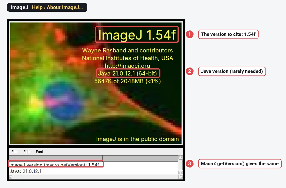
To cite ImageJ, cite **Schneider CA, Rasband WS, Eliceiri KW (2012). NIH Image to ImageJ: 25 years of image analysis. Nature Methods 9, 671–675. doi:10.1038/nmeth.2089, and give the version you used (Help › About ImageJ, for example 1.54f). If you used Fiji, cite Schindelin J et al. (2012). Fiji: an open-source platform for biological-image analysis. Nature Methods 9, 676–682. doi:10.1038/nmeth.2019**, and cite each plugin that has its own paper.
Which paper to cite
| You used | Cite | DOI |
|---|---|---|
| ImageJ 1.x (the download from imagej.net/ij) | Schneider, Rasband & Eliceiri 2012, Nature Methods | 10.1038/nmeth.2089 |
| Fiji | Schindelin et al. 2012, Nature Methods (and often the ImageJ paper too) | 10.1038/nmeth.2019 |
| ImageJ2 features (Ops, SciJava scripts, the ImageJ2 data model) | Rueden et al. 2017, BMC Bioinformatics | 10.1186/s12859-017-1934-z |
| A plugin with its own paper | That paper as well (see below) |
If you are unsure whether you used ImageJ or Fiji: Fiji's main window is titled "(Fiji Is Just) ImageJ" and its Plugins menu is long. When in doubt, cite both Schindelin et al. and Schneider et al.; it costs one line.
Some journals want a software reference in addition to, or instead of, a paper. The form ImageJ's author has long asked for is: Rasband WS. ImageJ. U.S. National Institutes of Health, Bethesda, Maryland, USA. https://imagej.net/ij/, 1997–2018. (Older papers give the address as imagej.nih.gov/ij, which now redirects.)
How to find the version you used
- Help › About ImageJ shows it in large type: here, ImageJ 1.54f.
- In Fiji, click the status bar under the toolbar: it shows a pair of numbers such as 2.14.0/1.54f, the ImageJ2 version and then the ImageJ 1.x version. Quote both.
- In a macro,
getVersion()returns the same string, handy for logging it with your results. - For a plugin, look at its documentation or About entry, or at the version in its .jar file name in Fiji's plugins or jars folder.
Record it when you analyse, not when you write the paper: Fiji updates itself, and a year later the version on your computer is not the one that made your figures.
Citing plugins
Name every plugin that did real work, with its version, and cite its paper. Some common ones:
| Plugin | Cite | DOI |
|---|---|---|
| TrackMate | Tinevez et al. 2017, Methods; for TrackMate 7 and later, Ershov et al. 2022, Nature Methods | 10.1016/j.ymeth.2016.09.016; 10.1038/s41592-022-01507-1 |
| Bio-Formats (opening .czi, .nd2, .lif) | Linkert et al. 2010, Journal of Cell Biology | 10.1083/jcb.201004104 |
| JACoP (colocalisation) | Bolte & Cordelières 2006, Journal of Microscopy | 10.1111/j.1365-2818.2006.01706.x |
| Trainable Weka Segmentation | Arganda-Carreras et al. 2017, Bioinformatics | 10.1093/bioinformatics/btx180 |
| Colour Deconvolution 2 | Landini, Martinelli & Piccinini 2021, Bioinformatics; and the method, Ruifrok & Johnston 2001 | 10.1093/bioinformatics/btaa847 |
| Cellpose (via its Fiji wrapper or Python) | Stringer et al. 2021, Nature Methods | 10.1038/s41592-020-01018-x |
Built-in commands (Analyze Particles, Subtract Background, the automatic threshold methods) do not need a separate citation, but name the method: "Otsu threshold", "rolling-ball background subtraction, radius 50 pixels". Where a method has a paper, such as Otsu's threshold (Otsu 1979), citing it is good practice.
Methods sentences to copy
Fill in the parts in brackets. Each one names the software and version, the steps and the settings, which is what a reviewer needs to repeat the analysis.
Cell counting
Nuclei were counted in [Fiji (ImageJ 1.54f; Schindelin et al. 2012)] on [DAPI] images: [Otsu] threshold, Fill Holes and Watershed, then Analyze Particles with a minimum size of [7 µm²]. [n] fields per [well] were analysed.
Fluorescence intensity (CTCF)
Fluorescence was measured in [ImageJ 1.54f (Schneider et al. 2012)] on the original [16-bit] images. Corrected total cell fluorescence was calculated as integrated density − (cell area × mean background of three adjacent regions).
Western blot densitometry
Band intensities were quantified in [ImageJ 1.54f (Schneider et al. 2012)] with the Gels tool (Analyze › Gels) on unsaturated [16-bit] images; each band was normalised to [total protein / β-actin] in the same lane and expressed relative to [control].
Particle or grain size
Particle sizes were measured in [ImageJ 1.54f (Schneider et al. 2012)] on [TEM] images calibrated from the embedded pixel size ([0.25] nm per pixel): [Default] threshold, Watershed, and Analyze Particles excluding particles touching the image edge; the equivalent circular diameter was calculated from each area. [n] particles from [3] images were measured.
More detail on each: counting cells, fluorescence intensity and CTCF, western blot quantification, grain size from SEM.
Reference-manager formats
APA (7th edition)
Schneider, C. A., Rasband, W. S., & Eliceiri, K. W. (2012). NIH Image to ImageJ: 25 years of image analysis. Nature Methods, 9(7), 671–675. https://doi.org/10.1038/nmeth.2089
Schindelin, J., Arganda-Carreras, I., Frise, E., Kaynig, V., Longair, M., Pietzsch, T., Preibisch, S., Rueden, C., Saalfeld, S., Schmid, B., Tinevez, J.-Y., White, D. J., Hartenstein, V., Eliceiri, K., Tomancak, P., & Cardona, A. (2012). Fiji: An open-source platform for biological-image analysis. Nature Methods, 9(7), 676–682. https://doi.org/10.1038/nmeth.2019
Rueden, C. T., Schindelin, J., Hiner, M. C., DeZonia, B. E., Walter, A. E., Arena, E. T., & Eliceiri, K. W. (2017). ImageJ2: ImageJ for the next generation of scientific image data. BMC Bioinformatics, 18(1), 529. https://doi.org/10.1186/s12859-017-1934-z
Vancouver
Schneider CA, Rasband WS, Eliceiri KW. NIH Image to ImageJ: 25 years of image analysis. Nat Methods. 2012;9(7):671-5. doi:10.1038/nmeth.2089
Schindelin J, Arganda-Carreras I, Frise E, Kaynig V, Longair M, Pietzsch T, et al. Fiji: an open-source platform for biological-image analysis. Nat Methods. 2012;9(7):676-82. doi:10.1038/nmeth.2019
Rueden CT, Schindelin J, Hiner MC, DeZonia BE, Walter AE, Arena ET, et al. ImageJ2: ImageJ for the next generation of scientific image data. BMC Bioinformatics. 2017;18(1):529. doi:10.1186/s12859-017-1934-z
BibTeX
@article{schneider2012imagej,
  author  = {Schneider, Caroline A. and Rasband, Wayne S. and Eliceiri, Kevin W.},
  title   = {{NIH} Image to {ImageJ}: 25 years of image analysis},
  journal = {Nature Methods},
  year    = {2012},
  volume  = {9},
  number  = {7},
  pages   = {671--675},
  doi     = {10.1038/nmeth.2089}
}

@article{schindelin2012fiji,
  author  = {Schindelin, Johannes and Arganda-Carreras, Ignacio and Frise, Erwin and Kaynig, Verena and Longair, Mark and Pietzsch, Tobias and Preibisch, Stephan and Rueden, Curtis and Saalfeld, Stephan and Schmid, Benjamin and Tinevez, Jean-Yves and White, Daniel James and Hartenstein, Volker and Eliceiri, Kevin and Tomancak, Pavel and Cardona, Albert},
  title   = {{Fiji}: an open-source platform for biological-image analysis},
  journal = {Nature Methods},
  year    = {2012},
  volume  = {9},
  number  = {7},
  pages   = {676--682},
  doi     = {10.1038/nmeth.2019}
}

@article{rueden2017imagej2,
  author  = {Rueden, Curtis T. and Schindelin, Johannes and Hiner, Mark C. and DeZonia, Barry E. and Walter, Alison E. and Arena, Ellen T. and Eliceiri, Kevin W.},
  title   = {{ImageJ2}: {ImageJ} for the next generation of scientific image data},
  journal = {BMC Bioinformatics},
  year    = {2017},
  volume  = {18},
  number  = {1},
  pages   = {529},
  doi     = {10.1186/s12859-017-1934-z}
}
To import into Zotero, Mendeley or EndNote, paste the DOI into the tool's "add by identifier" box; it fetches the full record.
An easier way: a methods sentence written for you in LabCanvas
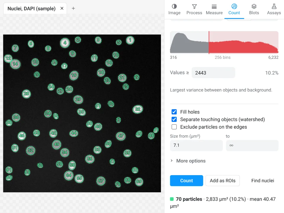
Writing the methods is the slow part of citing image analysis: the version, the plugin, the threshold, the sizes. LabCanvas Image Analysis, which runs in the browser with nothing to install, writes that sentence for you with the settings filled in.
- Open your image in Image Analysis and measure it, for example Count › Count particles.
- Under the results table, copy the Methods sentence: it names the threshold method, the size limits, the background step and the pixel size you used.
- Paste it after the software citation. LabCanvas has no paper or DOI, so name it, its address and when you used it:
Images were analysed in LabCanvas Image Analysis (www.labcanvas.com, accessed [month year]).
Why it is a good choice when you have to write the methods:
- The settings are in the sentence, so nothing is left out or misremembered months later.
- ImageJ's names and definitions for the measurements (Area, Mean, IntDen, RawIntDen and the rest); saying so in the methods helps readers compare numbers with ImageJ.
- Checked measurements: if a reviewer asks, the validation page lists more than 4,000 automated checks against scikit-image, SciPy and NumPy.
- Free with a LabCanvas account; nothing to install, on any computer or tablet.
Open Image Analysis, or read the complete guide.
ImageJ and Fiji are independent open-source projects; LabCanvas is not affiliated with them.
References
- Rueden CT, Schindelin J, Hiner MC, DeZonia BE, Walter AE, Arena ET, Eliceiri KW (2017). ImageJ2: ImageJ for the next generation of scientific image data. BMC Bioinformatics 18, 529. doi:10.1186/s12859-017-1934-z
- Schindelin J, Arganda-Carreras I, Frise E, et al. (2012). Fiji: an open-source platform for biological-image analysis. Nature Methods 9, 676–682. doi:10.1038/nmeth.2019
- Schneider CA, Rasband WS, Eliceiri KW (2012). NIH Image to ImageJ: 25 years of image analysis. Nature Methods 9, 671–675. doi:10.1038/nmeth.2089
Frequently asked questions
How do I cite ImageJ?
Cite Schneider CA, Rasband WS, Eliceiri KW (2012). NIH Image to ImageJ: 25 years of image analysis. Nature Methods 9, 671–675, doi:10.1038/nmeth.2089, and give the version you used (Help › About ImageJ), for example ImageJ 1.54f.
How do I cite Fiji?
Cite Schindelin J et al. (2012). Fiji: an open-source platform for biological-image analysis. Nature Methods 9, 676–682, doi:10.1038/nmeth.2019. Fiji runs ImageJ, so many authors also cite the ImageJ paper (Schneider et al. 2012), and every plugin you used that has its own paper.
What is the difference between citing ImageJ, Fiji and ImageJ2?
ImageJ (ImageJ 1.x) is Wayne Rasband’s original program: cite Schneider et al. 2012. Fiji is a distribution of ImageJ with many plugins: cite Schindelin et al. 2012. ImageJ2 is the rewritten core that Fiji runs on: cite Rueden et al. 2017 if you used its features (for example scripts with ImageJ Ops or the SciJava framework).
Where do I find my ImageJ version?
Help › About ImageJ shows the version in large type (for example ImageJ 1.54f). In Fiji, clicking the status bar of the main window also shows it, as a pair such as 2.14.0/1.54f: the ImageJ2 version, then the ImageJ 1.x version. In a macro, getVersion() returns it.
Do I need to cite plugins separately?
Yes, when the plugin has its own publication: TrackMate, Bio-Formats, JACoP, Trainable Weka Segmentation, Colour Deconvolution and many others ask to be cited. The plugin’s documentation page usually names the paper. Name the plugin and its version in the methods too.
Try it in LabCanvas
Every app is free to start in your browser. Create an account and begin.
Start Image Analysis free in your browserKeep reading

ImageJ vs LabCanvas Image Analysis: an honest comparison
ImageJ/Fiji or LabCanvas Image Analysis? Six everyday jobs done in both, six newer ones (macros, vendor files, 3D, StarDist, big images) and which to use.

How to use ImageJ: a beginner's tutorial for the 10 everyday jobs (2026)
How to use ImageJ and Fiji: get started, open an image, then 10 everyday jobs (scale, measure, threshold, count, background, blots) with exact menu paths.

How to set the scale and add a scale bar in ImageJ (with a calibration image to practise on)
Set the scale in ImageJ (Analyze › Set Scale) from a micrometer, scale bar or pixel size, add a scale bar, and practise on a free calibration image.