Open DICOM files in ImageJ (and a series as a stack)
Open DICOM files in ImageJ with File › Open, a whole series as a stack with Import › Image Sequence, read the tags, measure HU, and open compressed DICOM.
Contents
To open a DICOM file in ImageJ, use File › Open… (or drag the file onto the ImageJ window): ImageJ 1.x and Fiji read uncompressed DICOM directly, with the pixel spacing in mm and CT values in Hounsfield units. To open a whole series as one stack, put its slices in a folder and use File › Import › Image Sequence…. Image › Show Info lists the DICOM tags. Compressed DICOM (JPEG, JPEG 2000, RLE) needs Fiji's File › Import › Bio-Formats.
What you need: ImageJ 1.x or Fiji (Fiji for compressed files). Uncompressed DICOM files; one folder per series. The example is a synthetic CT-like phantom written for this guide, not a patient: 12 slices of 256 × 256 pixels, 0.7 mm pixels, 2.5 mm slices, plus one slice saved with RLE compression. ImageJ's own sample, File › Open Samples › CT (16-bit DICOM), also works (it downloads from the internet). Time: 10 minutes.
ImageJ is research software. It is fine for measuring images in a study, but it is not a certified medical device: do not use it for diagnosis, and anonymise patient data before you share files.
Open one DICOM file, step by step
Step 1: Open the file
Choose File › Open… and pick the .dcm file (DICOM files without an extension open too). The title bar shows the size in mm: ImageJ took the pixel spacing from the file. The image is shown with the file's own window (here centre 40, width 400, so the display runs from -160 to 240).
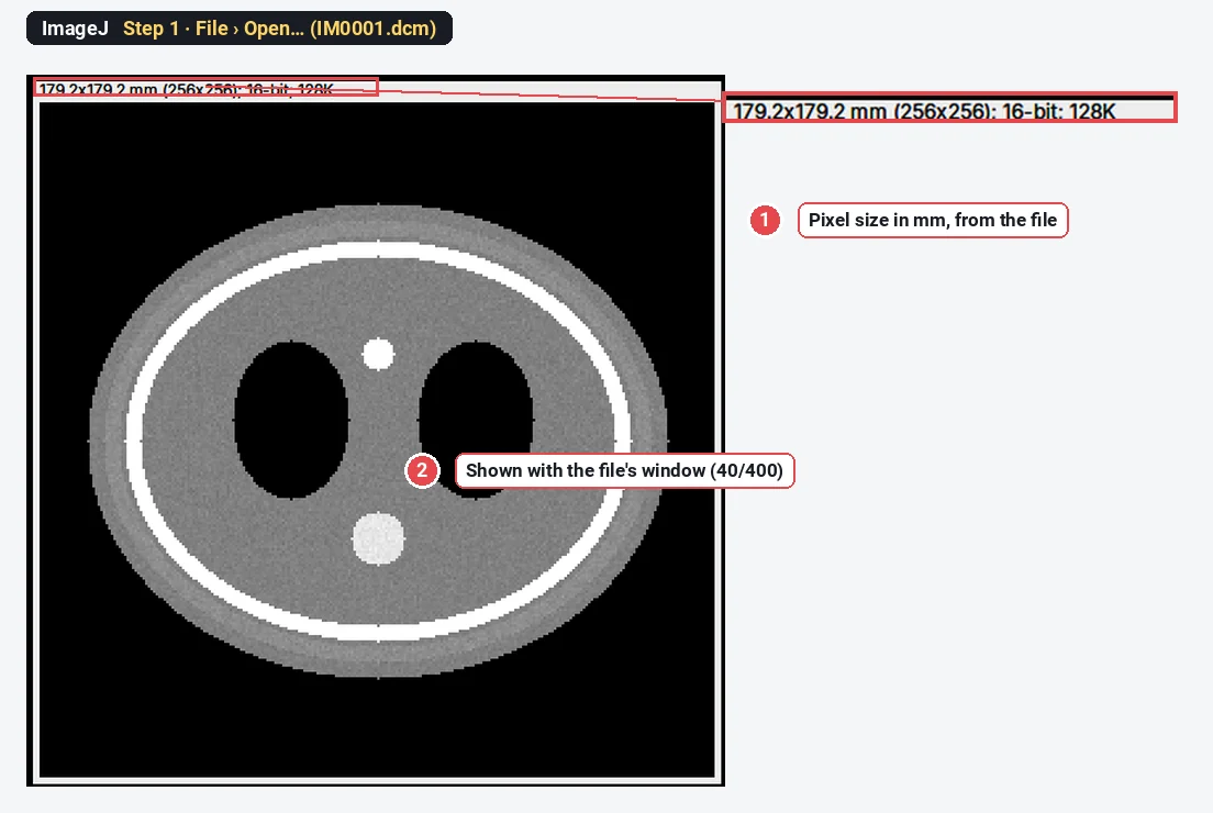
ImageJ also applies the Rescale Slope and Rescale Intercept from the header as a calibration. The stored pixel in the centre is 1067; ImageJ shows and measures it as 1067 − 1024 = 43 HU.
Step 2: Read the tags with Show Info
Choose Image › Show Info… (Ctrl I). Each line is a tag: group and element number, name, value.
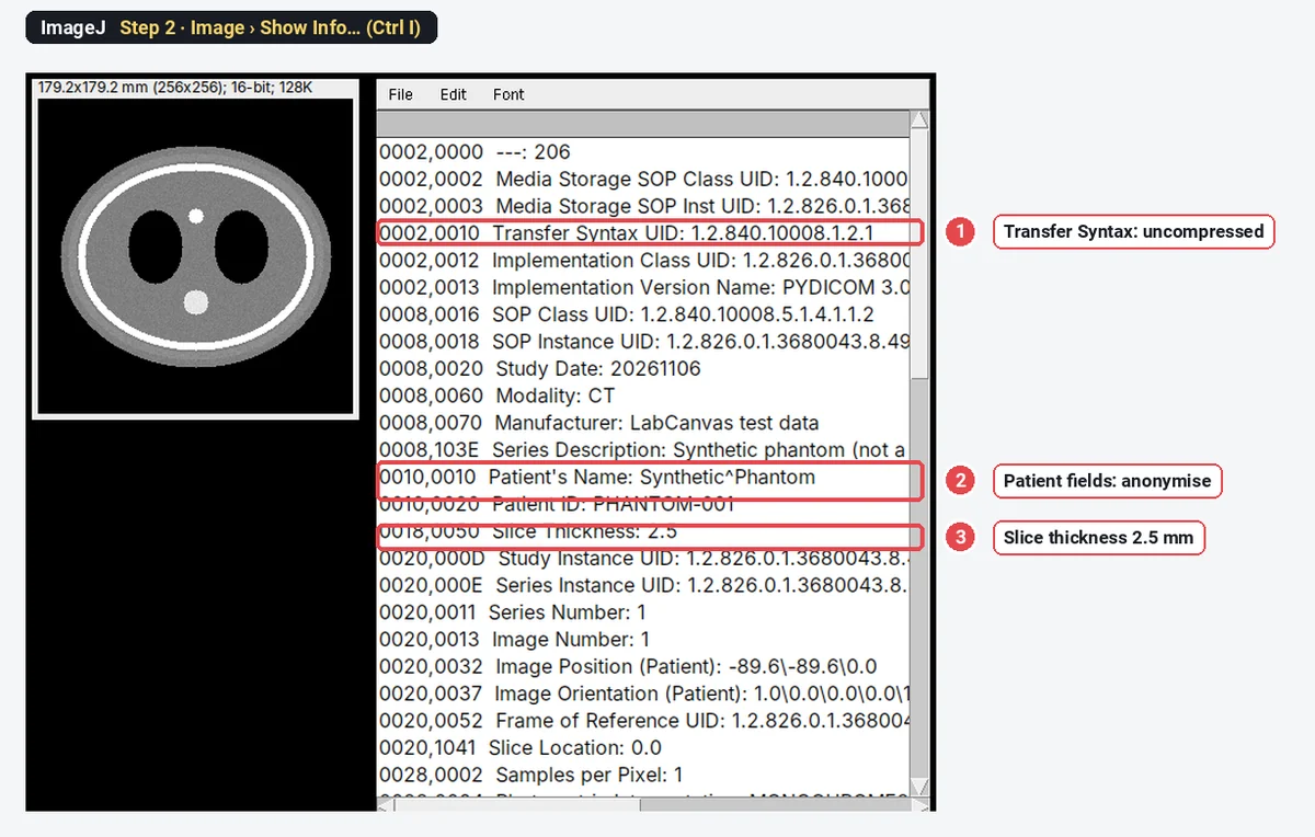
| Tag | What it tells you |
|---|---|
| 0002,0010 Transfer Syntax UID | Whether the pixels are compressed (see below) |
| 0008,0060 Modality | CT, MR, US, and so on |
| 0010,0010 and 0010,0020 Patient's Name, Patient ID | Personal data: remove before sharing |
| 0018,0050 Slice Thickness | Slice thickness in mm |
| 0020,0013 Image Number | The slice's place in the series |
| 0028,0030 Pixel Spacing | Pixel size in mm (row, column) |
| 0028,1050 and 0028,1051 Window Center, Window Width | The display window |
| 0028,1052 and 0028,1053 Rescale Intercept, Rescale Slope | Stored value to real value (HU for CT) |
From a macro, getInfo("0028,0030") returns a single tag's value.
Open a DICOM series as a stack
A CT or MR series is usually one file per slice. ImageJ opens them as one stack.
Step 3: Import the folder
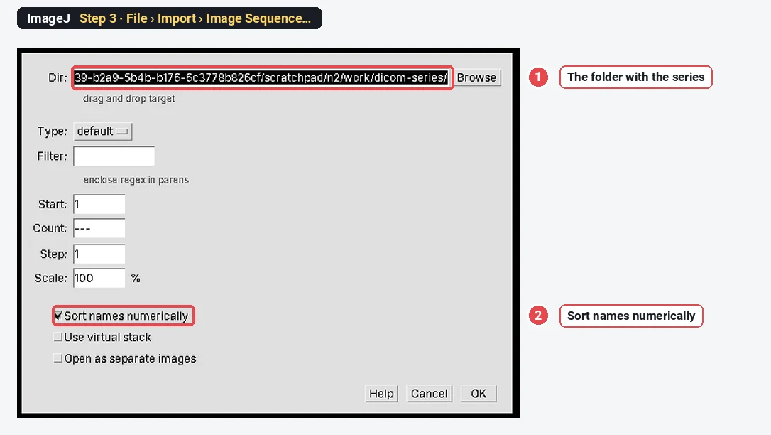
Put the slices of one series in their own folder. Choose File › Import › Image Sequence…, browse to the folder, keep Sort names numerically ticked, and click OK.
Step 4: Check the stack
The series opens as one window with a slider. The title shows the slice number and the file it came from. Image › Properties… shows the voxel size: here 0.7 × 0.7 × 2.5 mm, taken from the files, so Image › Stacks › Orthogonal Views and 3D tools have the right proportions.
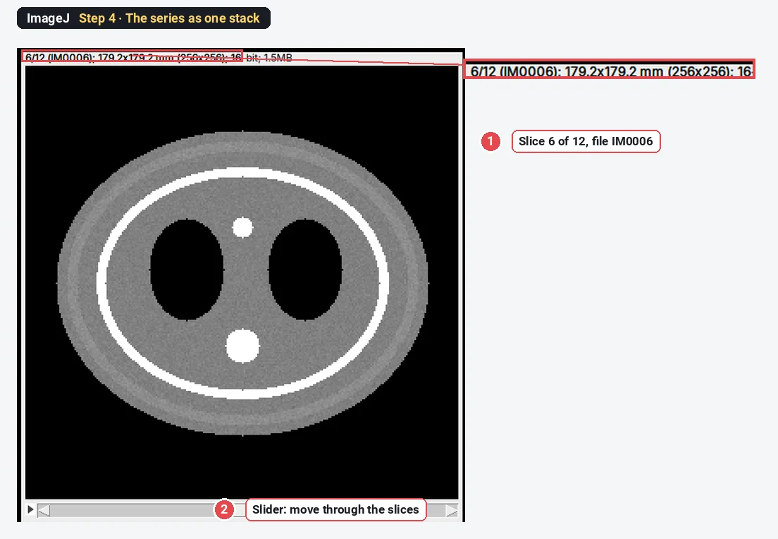
If the slices come in the wrong order (files named by acquisition time, or several series mixed), check that the folder holds only one series, then sort by Image Number or Slice Location, which Show Info lists for each slice.
Step 5: Measure Hounsfield units
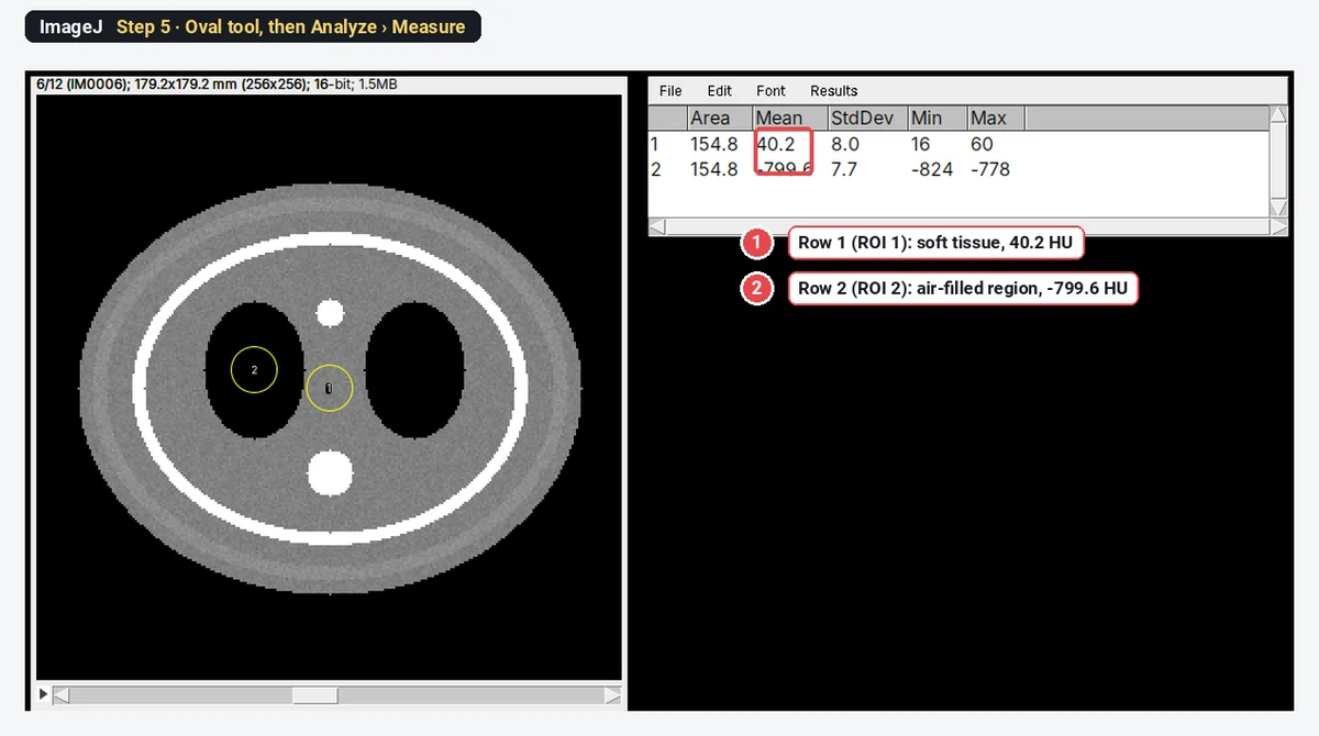
Draw an Oval in the tissue you want, choose Analyze › Set Measurements… (Area, Mean, Standard deviation, Min & max), and press M (Analyze › Measure). Because ImageJ applied the rescale values, Mean is in HU and Area in mm².
The phantom was written with 40 HU of soft tissue and -800 HU in the air-filled regions, with noise of 8 HU, which is what ImageJ reads back.
Why won't ImageJ open my compressed DICOM file?
Many scanners and PACS exports compress the pixel data (JPEG, JPEG 2000 or RLE). The DICOM reader built into ImageJ 1.x reads only uncompressed files, and says so:
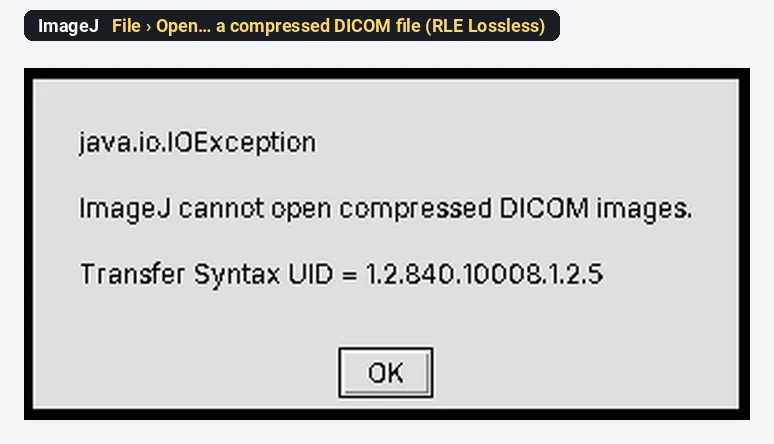
The Transfer Syntax UID tells you which compression it is:
| Transfer Syntax UID | Meaning | ImageJ 1.x File › Open |
|---|---|---|
| 1.2.840.10008.1.2 | Implicit VR Little Endian, uncompressed | Opens |
| 1.2.840.10008.1.2.1 | Explicit VR Little Endian, uncompressed | Opens |
| 1.2.840.10008.1.2.4.50 | JPEG baseline | No |
| 1.2.840.10008.1.2.4.70 | JPEG lossless | No |
| 1.2.840.10008.1.2.4.90 and .91 | JPEG 2000 | No |
| 1.2.840.10008.1.2.5 | RLE lossless | No |
In Fiji, open it with File › Import › Bio-Formats: the Bio-Formats DICOM reader handles uncompressed data, baseline JPEG, JPEG 2000 (lossy and lossless) and RLE lossless (Linkert et al. 2010). Choose View stack with: Hyperstack and tick Group files with similar names to open a series. If a file still does not open (for example JPEG lossless, which is not in that list), decompress it with a DICOM tool, such as dcmdjpeg from DCMTK or pydicom in Python, and open the result in ImageJ.
DICOM options worth knowing
Edit › Options › DICOM… changes how ImageJ 1.x opens DICOM:
- Open as 32-bit float: the image is converted to real values (HU for CT) when it opens, instead of keeping the stored 16-bit values with a calibration.
- Ignore rescale slope: uses the stored values without the slope.
To save the images for other software, use File › Save As › Tiff…. ImageJ 1.x cannot write DICOM. If the other program should see Hounsfield units, convert first with Image › Type › 32-bit: the stored 1067 becomes 43. A 16-bit TIFF keeps the stored values (HU + 1024 here), and only ImageJ knows the calibration.
Is ImageJ good for medical imaging?
For research, yes, within limits:
- Good at: reading uncompressed DICOM, keeping pixel spacing and HU, ROI measurements on slices and stacks, projections, orthogonal views, macros for many files; Fiji adds Bio-Formats for compressed files, 3D viewing and segmentation plugins.
- Not for: diagnosis or clinical reports (it is not certified medical software), connecting to a PACS, writing DICOM, or advanced volume rendering and registration, where dedicated research tools are better suited.
Pitfalls
- Several series in one folder. A scout image or a second series ends up in the stack. One series per folder.
- Compressed files. Check the Transfer Syntax UID in Show Info before you blame the file.
- Raw values outside ImageJ. A 16-bit TIFF holds stored values; other software will not apply the rescale. Convert to 32-bit for real units.
- Window changes are only display. Changing brightness and contrast (or the stored window) does not change measurements.
- Uneven slice spacing. Gaps or overlapping slices make the voxel depth wrong; check Slice Location for every slice.
- Patient data. DICOM headers carry names, dates and IDs. Anonymise before sharing, and do not post real headers.
Reporting it
DICOM images were opened in ImageJ [1.54f] (Schneider et al. 2012) as stacks (File › Import › Image Sequence; voxel size [0.7 × 0.7 × 2.5] mm from the headers). Mean attenuation was measured in [circular ROIs of 154.8 mm²] in [tissue] and reported in Hounsfield units.
Switch to LabCanvas: measure the exported TIFF
LabCanvas Image Analysis does not read DICOM. If your measurement is a simple one (areas, mean values, counts) on images you have exported as TIFF, it runs in the browser with nothing to install.
- In ImageJ, convert to 32-bit and File › Save As › Tiff…, so the file holds the real values.
- In LabCanvas, click Open an image and choose the TIFF; check the scale in the Image tab (set it with Scale if the pixel size did not come across).
- Draw ROIs and click Measure in the Measure tab (Area, Mean, StdDev, Min and Max).
- Send to Graphs to compare groups.
Why switch for this job:
- No install, ImageJ's names for the measurements, on any computer.
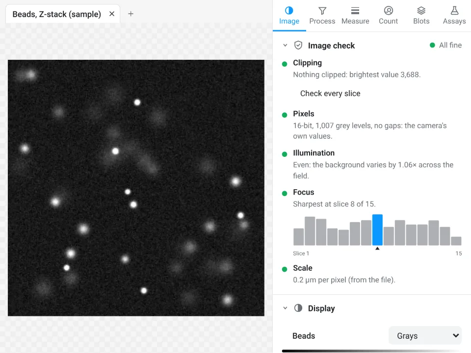
- An image check before you measure: clipping and the number of grey levels are flagged, so you notice an 8-bit export that lost the HU range.
- Results straight to Graphs with statistics, and a methods sentence to copy.
For DICOM itself (opening series, headers, compressed files, 3D), ImageJ and Fiji are the right tools.
Open Image Analysis: free with a LabCanvas account (downloading files is part of LabCanvas Pro). ImageJ and Fiji are independent open-source projects; LabCanvas is not affiliated with them.
References
- Linkert M, Rueden CT, Allan C, et al. (2010). Metadata matters: access to image data in the real world. Journal of Cell Biology 189, 777–782. doi:10.1083/jcb.201004104
- Schindelin J, Arganda-Carreras I, Frise E, et al. (2012). Fiji: an open-source platform for biological-image analysis. Nature Methods 9, 676–682. doi:10.1038/nmeth.2019
- Schneider CA, Rasband WS, Eliceiri KW (2012). NIH Image to ImageJ: 25 years of image analysis. Nature Methods 9, 671–675. doi:10.1038/nmeth.2089
Frequently asked questions
Can ImageJ open DICOM files?
Yes. File › Open (or drag and drop) opens uncompressed DICOM files in ImageJ 1.x and Fiji, with the pixel size in mm and the Rescale Slope and Intercept applied, so CT values read in Hounsfield units. File › Import › Image Sequence opens a folder of slices as one stack.
Why does ImageJ say it cannot open compressed DICOM images?
The built-in DICOM reader of ImageJ 1.x reads only uncompressed data. A JPEG, JPEG 2000 or RLE compressed file gives "ImageJ cannot open compressed DICOM images" with its Transfer Syntax UID. Open it in Fiji with File › Import › Bio-Formats, which reads baseline JPEG, JPEG 2000 and RLE lossless DICOM, or decompress it first with a DICOM tool.
Is ImageJ good for medical imaging?
For research, yes: it reads uncompressed DICOM, keeps the pixel spacing and Hounsfield calibration, measures ROIs and handles stacks, and Fiji adds 3D tools. It is not certified medical software, so it is not for diagnosis or clinical reporting, and it has no PACS connection or DICOM saving.
How do I see the DICOM header in ImageJ?
Image › Show Info (Ctrl I) lists every tag with its group and element number, name and value: patient fields, modality, pixel spacing, slice thickness, window centre and width, rescale values and the Transfer Syntax UID.
How do I open a DICOM series as a 3D stack?
Put the slices of one series in their own folder and use File › Import › Image Sequence with Sort names numerically ticked. ImageJ reads the slice spacing, so Image › Properties shows the voxel depth (2.5 mm in the example) and Orthogonal Views and 3D tools have the right proportions.
How do I export DICOM to TIFF?
Open the file or series, convert with Image › Type › 32-bit if you want the Hounsfield values themselves in the file (16-bit keeps the raw stored values), then File › Save As › Tiff. ImageJ 1.x cannot save DICOM.
Try it in LabCanvas
Every app is free to start in your browser. Create an account and begin.
Start Image Analysis free in your browserKeep reading

ImageJ vs LabCanvas Image Analysis: an honest comparison
ImageJ/Fiji or LabCanvas Image Analysis? Six everyday jobs done in both, six newer ones (macros, vendor files, 3D, StarDist, big images) and which to use.

How to use ImageJ: a beginner's tutorial for the 10 everyday jobs (2026)
How to use ImageJ and Fiji: get started, open an image, then 10 everyday jobs (scale, measure, threshold, count, background, blots) with exact menu paths.

How to set the scale and add a scale bar in ImageJ (with a calibration image to practise on)
Set the scale in ImageJ (Analyze › Set Scale) from a micrometer, scale bar or pixel size, add a scale bar, and practise on a free calibration image.